

5.6 PSF Variations with Field Position
The WFPC2 PSFs vary with field position due to field-dependent aberrations, obscuration shifting, and scattering. This complicates photometry, PSF subtraction, and deconvolution (Krist, 1995).
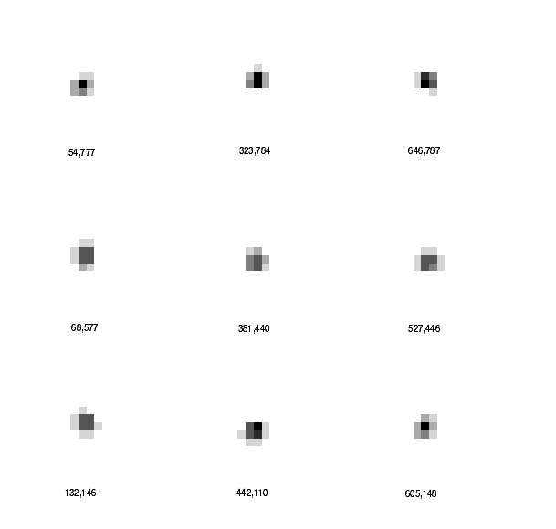
The coma and astigmatism aberrations vary significantly within a camera across the field-of-view. These variations are simply part of the optical design. At the extreme corners of the WFC CCDs, away from the OTA axis, there is about 1/5 wave of astigmatism (referenced at 633 nm), which decreases to nearly zero at the CCD centers. Astigmatism at this level causes the PSF core to become elliptical and slightly less sharp; note the flattening of the PSF at pixel positions (54,777) and (605,148) in Figure 5.5. Coma also varies, but to a much lesser extent. Coma and astigmatism variations are considerably smaller in PC1 (though we note the astigmatism at the center of PC1 is fairly significant - see Table 5.2).
Table 5.3: PC Point Spread Functions. Shown as percentages (out of 100 percent) of the total flux in a 5 by 5 pixel region. On the left in each case is a model PSF with the observed wavefront errors and pixel response function. On the right is the diffraction limited case for comparison.
| WFPC2 Model PSF |
|
Diffraction Limited PSF |
| 2000 Å: Peak near corner of PC pixel |
| 0.9 |
2.3 |
2.3 |
0.9 |
0.3 |
|
0.2 |
0.6 |
0.5 |
0.3 |
0.1 |
| 2.5 |
10.3 |
12.7 |
2.5 |
0.5 |
|
0.6 |
17.6 |
20.9 |
0.5 |
0.1 |
| 1.9 |
11.2 |
13.3 |
2.6 |
0.5 |
|
0.5 |
20.9 |
26.0 |
0.6 |
0.2 |
| 0.9 |
2.1 |
2.7 |
1.4 |
0.3 |
|
0.3 |
0.5 |
0.6 |
0.3 |
0.1 |
| 0.3 |
0.5 |
0.5 |
0.4 |
0.2 |
|
0.1 |
0.1 |
0.2 |
0.1 |
0.2 |
| Peak near center of pixel |
| 0.3 |
0.7 |
1.3 |
0.8 |
0.5 |
|
0.1 |
0.3 |
0.4 |
0.2 |
0.2 |
| 0.7 |
2.6 |
6.4 |
3.2 |
0.8 |
|
0.3 |
0.6 |
4.9 |
0.6 |
0.3 |
| 1.2 |
6.3 |
25.0 |
6.9 |
1.4 |
|
0.4 |
4.9 |
62.9 |
6.3 |
0.4 |
| 0.6 |
2.2 |
5.9 |
3.6 |
0.9 |
|
0.3 |
0.5 |
6.3 |
0.7 |
0.4 |
| 0.4 |
0.8 |
1.4 |
1.1 |
0.4 |
|
0.2 |
0.3 |
0.4 |
0.4 |
0.2 |
| 4000 Å: Peak near corner of PC pixel |
| 0.9 |
2.5 |
3.5 |
1.2 |
0.2 |
|
0.3 |
2.5 |
2.5 |
0.3 |
0.1 |
| 3.6 |
10.8 |
12.0 |
3.1 |
0.4 |
|
2.5 |
14.3 |
15.8 |
2.7 |
0.2 |
| 2.7 |
11.5 |
12.9 |
3.5 |
0.4 |
|
2.5 |
15.8 |
17.6 |
3.0 |
0.2 |
| 0.8 |
3.0 |
3.4 |
1.3 |
0.3 |
|
0.3 |
2.7 |
3.0 |
0.4 |
0.1 |
| 0.2 |
0.4 |
0.4 |
0.3 |
0.2 |
|
0.1 |
0.2 |
0.2 |
0.1 |
0.1 |
| Peak near center of pixel |
| 0.3 |
0.6 |
0.8 |
0.9 |
0.3 |
|
0.1 |
0.2 |
0.3 |
0.2 |
0.1 |
| 0.9 |
4.0 |
6.6 |
4.9 |
0.8 |
|
0.2 |
3.8 |
4.4 |
3.8 |
0.2 |
| 0.9 |
6.9 |
26.1 |
7.0 |
0.8 |
|
0.3 |
4.5 |
49.4 |
5.1 |
0.4 |
| 0.5 |
3.4 |
6.8 |
4.9 |
0.9 |
|
0.2 |
3.8 |
5.0 |
4.3 |
0.2 |
| 0.2 |
0.6 |
0.8 |
0.9 |
0.4 |
|
0.1 |
0.2 |
0.4 |
0.2 |
0.1 |
| 6000 Å: Peak near corner of PC pixel |
| 2.0 |
2.6 |
3.4 |
2.4 |
0.5 |
|
2.1 |
2.3 |
2.0 |
2.1 |
0.2 |
| 3.4 |
9.8 |
10.4 |
2.9 |
0.6 |
|
2.2 |
11.4 |
12.8 |
2.1 |
0.6 |
| 2.8 |
10.6 |
11.2 |
3.2 |
0.6 |
|
2.0 |
12.9 |
14.1 |
2.1 |
0.6 |
| 1.6 |
2.8 |
3.1 |
2.4 |
0.5 |
|
2.0 |
2.0 |
2.1 |
2.3 |
0.2 |
| 0.4 |
0.6 |
0.7 |
0.5 |
0.2 |
|
0.2 |
0.5 |
0.6 |
0.2 |
0.1 |
| Peak near center of pixel |
| 0.5 |
1.2 |
1.7 |
1.7 |
0.6 |
|
0.2 |
1.5 |
1.8 |
1.4 |
0.3 |
| 1.7 |
3.1 |
5.9 |
3.6 |
1.6 |
|
1.5 |
2.4 |
4.3 |
2.2 |
1.6 |
| 2.0 |
6.0 |
20.7 |
6.6 |
1.8 |
|
1.8 |
4.4 |
31.6 |
5.3 |
2.0 |
| 1.2 |
2.9 |
6.2 |
3.7 |
1.7 |
|
1.4 |
2.1 |
5.4 |
2.3 |
1.7 |
| 0.4 |
1.3 |
2.0 |
1.7 |
0.6 |
|
0.2 |
1.5 |
1.9 |
1.7 |
0.3 |
| 8000 Å: Peak near corner of PC pixel |
| 1.6 |
1.9 |
2.2 |
1.9 |
1.1 |
|
1.8 |
0.9 |
0.9 |
1.5 |
1.1 |
| 2.1 |
9.3 |
9.7 |
2.1 |
1.1 |
|
0.9 |
11.7 |
12.6 |
1.0 |
1.4 |
| 2.0 |
9.8 |
10.1 |
2.4 |
1.1 |
|
0.9 |
12.6 |
13.3 |
1.0 |
1.5 |
| 1.4 |
2.1 |
2.1 |
1.8 |
1.1 |
|
1.5 |
1.0 |
1.0 |
1.6 |
1.2 |
| 0.8 |
1.2 |
1.3 |
1.1 |
0.4 |
|
1.1 |
1.4 |
1.5 |
1.2 |
0.2 |
| Peak near center of pixel |
| 1.2 |
1.4 |
1.5 |
1.8 |
1.4 |
|
1.3 |
1.8 |
1.1 |
1.6 |
1.4 |
| 1.8 |
2.5 |
6.0 |
2.8 |
1.6 |
|
1.8 |
1.5 |
6.2 |
1.7 |
1.6 |
| 1.6 |
6.0 |
15.4 |
6.6 |
1.5 |
|
1.1 |
6.2 |
22.5 |
7.1 |
1.0 |
| 1.3 |
2.7 |
6.3 |
3.0 |
1.7 |
|
1.6 |
1.7 |
7.1 |
1.9 |
1.7 |
| 1.0 |
1.4 |
1.5 |
1.7 |
1.4 |
|
1.4 |
1.6 |
1.0 |
1.7 |
1.5 |
Table 5.4: WFC Point Spread Functions. Shown as percentages (out of 100 percent) of the total flux in a 5 by 5 pixel region. On the left in each case is a model PSF with the observed wavefront errors and pixel response function. On the right is the diffraction limited case for comparison.
| WFPC2 Model PSF |
|
Diffraction Limited PSF |
| 2000 Å: Peak near corner of WF pixel |
| 0.5 |
1.8 |
2.1 |
0.8 |
0.3 |
|
0.4 |
0.4 |
0.4 |
0.3 |
0.1 |
| 1.5 |
9.9 |
13.6 |
3.1 |
0.5 |
|
0.4 |
15.4 |
21.5 |
0.4 |
0.1 |
| 1.5 |
9.8 |
24.0 |
4.7 |
0.5 |
|
0.4 |
21.5 |
33.4 |
0.4 |
0.1 |
| 0.5 |
2.1 |
3.6 |
1.3 |
0.3 |
|
0.3 |
0.4 |
0.4 |
0.4 |
0.1 |
| 0.2 |
0.4 |
0.4 |
0.2 |
0.2 |
|
0.1 |
0.1 |
0.1 |
0.1 |
0.1 |
| Peak near center of pixel |
| 0.2 |
0.4 |
0.5 |
0.5 |
0.3 |
|
0.2 |
0.2 |
0.2 |
0.1 |
0.2 |
| 0.3 |
1.7 |
5.3 |
3.0 |
0.8 |
|
0.1 |
0.8 |
1.3 |
0.6 |
0.1 |
| 0.5 |
5.4 |
28.5 |
14.0 |
2.2 |
|
0.2 |
1.3 |
86.2 |
1.4 |
0.2 |
| 0.3 |
2.2 |
9.1 |
4.5 |
1.0 |
|
0.1 |
0.6 |
1.4 |
0.9 |
0.2 |
| 0.3 |
0.6 |
1.2 |
0.8 |
0.3 |
|
0.2 |
0.1 |
0.2 |
0.2 |
0.2 |
| 4000 Å: Peak near corner of WF pixel |
| 0.8 |
2.6 |
2.5 |
0.8 |
0.2 |
|
0.4 |
0.5 |
0.5 |
0.2 |
0.1 |
| 2.6 |
16.1 |
16.4 |
3.0 |
0.4 |
|
0.5 |
17.7 |
21.2 |
0.6 |
0.1 |
| 2.0 |
12.9 |
17.3 |
3.0 |
0.4 |
|
0.5 |
21.2 |
26.7 |
0.6 |
0.1 |
| 0.6 |
2.0 |
2.6 |
0.9 |
0.2 |
|
0.2 |
0.6 |
0.6 |
0.4 |
0.2 |
| 0.2 |
0.4 |
0.3 |
0.2 |
0.1 |
|
0.1 |
0.1 |
0.1 |
0.2 |
0.2 |
| Peak near center of pixel |
| 0.3 |
0.6 |
0.7 |
0.5 |
0.3 |
|
0.3 |
0.2 |
0.3 |
0.2 |
0.2 |
| 0.6 |
3.1 |
7.2 |
3.4 |
0.7 |
|
0.2 |
0.7 |
3.7 |
0.7 |
0.2 |
| 0.9 |
8.5 |
33.3 |
10.2 |
1.2 |
|
0.3 |
3.7 |
68.8 |
5.3 |
0.4 |
| 0.4 |
2.5 |
8.8 |
3.0 |
0.6 |
|
0.2 |
0.7 |
5.3 |
0.8 |
0.2 |
| 0.2 |
0.5 |
0.9 |
0.5 |
0.3 |
|
0.2 |
0.2 |
0.4 |
0.2 |
0.3 |
| 6000 Å: Peak near corner of WF pixel |
| 0.7 |
2.6 |
2.6 |
0.9 |
0.3 |
|
0.2 |
0.5 |
0.4 |
0.2 |
0.2 |
| 3.0 |
14.9 |
15.6 |
3.3 |
0.5 |
|
0.5 |
18.3 |
20.7 |
0.5 |
0.3 |
| 2.2 |
13.7 |
16.3 |
3.2 |
0.4 |
|
0.4 |
20.7 |
24.2 |
0.6 |
0.2 |
| 0.6 |
2.3 |
2.9 |
0.7 |
0.2 |
|
0.2 |
0.5 |
0.6 |
0.2 |
0.2 |
| 0.2 |
0.3 |
0.3 |
0.2 |
0.2 |
|
0.2 |
0.3 |
0.2 |
0.2 |
0.2 |
| Peak near center of pixel |
| 0.3 |
0.7 |
0.9 |
0.6 |
0.2 |
|
0.2 |
0.3 |
0.2 |
0.3 |
0.2 |
| 0.7 |
4.1 |
7.8 |
4.1 |
0.8 |
|
0.3 |
1.8 |
6.1 |
1.9 |
0.3 |
| 1.0 |
8.6 |
30.4 |
9.4 |
1.3 |
|
0.2 |
6.1 |
54.9 |
6.2 |
0.3 |
| 0.5 |
2.8 |
8.2 |
3.5 |
0.6 |
|
0.3 |
1.9 |
6.2 |
2.5 |
0.3 |
| 0.2 |
0.5 |
1.0 |
0.6 |
0.3 |
|
0.2 |
0.3 |
0.3 |
0.3 |
0.2 |
| 8000 Å: Peak near corner of WF pixel |
| 1.0 |
3.0 |
2.9 |
1.1 |
0.3 |
|
0.1 |
2.0 |
2.0 |
0.2 |
0.2 |
| 3.6 |
13.1 |
13.6 |
4.0 |
0.5 |
|
2.0 |
15.8 |
17.3 |
2.2 |
0.1 |
| 2.6 |
12.6 |
14.2 |
3.5 |
0.5 |
|
2.0 |
17.3 |
19.3 |
2.5 |
0.1 |
| 0.8 |
3.2 |
3.6 |
1.0 |
0.3 |
|
0.2 |
2.2 |
2.5 |
0.2 |
0.2 |
| 0.2 |
0.4 |
0.5 |
0.2 |
0.1 |
|
0.2 |
0.1 |
0.1 |
0.2 |
0.1 |
| Peak near center of pixel |
| 0.2 |
0.8 |
0.8 |
0.7 |
0.3 |
|
0.1 |
0.2 |
0.3 |
0.2 |
0.1 |
| 0.9 |
4.6 |
6.9 |
4.4 |
1.0 |
|
0.2 |
3.5 |
4.6 |
3.4 |
0.2 |
| 0.9 |
7.5 |
30.8 |
8.1 |
1.2 |
|
0.3 |
4.6 |
52.0 |
5.0 |
0.3 |
| 0.5 |
3.1 |
7.3 |
3.7 |
0.7 |
|
0.1 |
3.4 |
5.1 |
3.9 |
0.2 |
| 0.2 |
0.5 |
1.0 |
0.7 |
0.2 |
|
0.1 |
0.2 |
0.3 |
0.2 |
0.1 |
Figure 5.5: PSF Variations with Field Position - Aberrations. Nine observed PSFs (filter F814W) are shown from a widely spaced grid on WF3. CCD pixel positions are labeled. Note the flattening of the PSF in the (54,777) and (605,148) positions.
The obscuration patterns due to the camera optics (relay secondary mirror and spiders) appear to shift with respect to the OTA obscurations, depending on field position. The interacting diffraction patterns of the WFPC2 and OTA spiders cause ripples in the spider diffraction spikes, which vary with field position as the two spiders shift relative to each other. In Figure 5.6 the OTA spider is hidden behind the WFPC2 spider at the field center and hence the diffraction spikes there have a simple, smooth appearance (c.f. position 446,425). At the CCD corners, however, one or more vanes of the OTA spider move out from behind the WFPC2 spider, and the double set of obscurations causes a "beating" pattern in the diffraction spikes.
The spiders also interact with light diffracted from zonal errors in the OTA mirrors, causing streaks in the scattering halo which vary in position and intensity.
Figure 5.6: PSF Variations with Field Position - Obscuration Shifts. Five saturated PSFs observed in F814W are shown from a widely spaced grid on WF4. Note the changes in the spider diffraction spikes. CCD pixel positions are labeled. The vertical feature is caused by saturation and blooming (see Blooming).
Space Telescope Science Institute
http://www.stsci.edu
Voice: (410) 338-1082
help@stsci.edu
|